So, there was this woman who came to see me last year. I’ll call her Mrs. K. She must have been in her early fifties, very put together, the kind of person who’s used to handling things on her own. She sat down and told me, very calmly, that for six months she’d been feeling a weird numbness on the right side of her face. It would come and go. Her dentist said it wasn’t a tooth problem. Her GP said maybe it was sinus.Now finally when someone asked for a MRI, the result showed chordoma at the skull base. She reveals it like an ignorant voice but she was trembling.
I notice things like that. When she finished, she just looked at me and said, “I don’t want to die from something I can’t even pronounce.”
I think of her sometimes when I’m explaining chordomas to new patients. Because the fear underneath the calm exterior, that’s the real thing I’m treating in those first few minutes. One of the honest things about who is sitting in front of me is that the outlook is not what it used to be.
Nothing. A Skull Base Surgeon in Gurgaon can now do things that would have sounded borderline impossible when I was still learning this craft.
Okay, so what even is a chordoma?
I find that when people understand something, even a little, the terror shrinks. It doesn’t disappear. But it becomes manageable.So let us walk through without any rush or tension, the way we talk with a cup of coffee.
When a human is in an embryo form, the spine is not fully formed, in place, it has a flexible little rod-like structure known as notochord, which functions as a scaffolding. Once the spine built itself around it, the scaffolding was supposed to dissolve and vanish. And in almost everybody, it does. But in roughly one out of a million people, a tiny pocket of those cells just doesn’t get the memo. It hangs around, quiet, doing nothing, for decades. Then one day it starts dividing. That lump is a chordoma. The Chordoma Foundation explains this better than I can, if you want to read more.
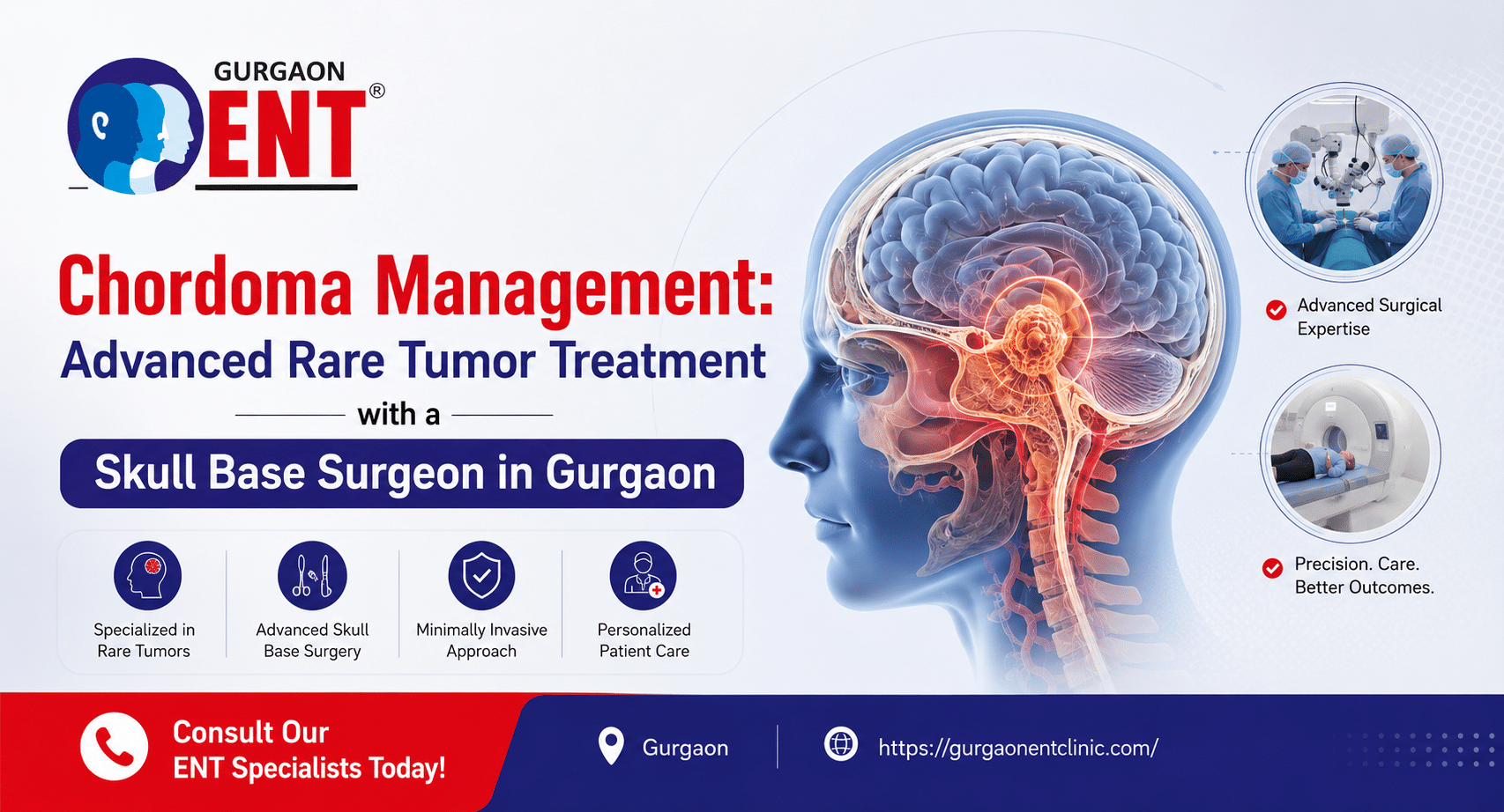
When a chordoma grows at the base of the skull, in a bone called the clivus, which sits right behind the nasal passages and right in front of the brainstem, the location is the whole problem. It’s not a big, open space. It’s a cramped little crossroads with major nerves running through it. Nerves for eye movement, facial sensation, swallowing, voice. The tumor pushes on these things very slowly. So the symptoms creep in.Symptoms like a patch on one of the cheeks, double vision when looking somewhere indirectly from the side at night, sudden nasal tone like the one when someone has a cold. Nothing that makes you call an ambulance. That’s exactly why diagnosis often comes late.
Why you need a real specialist for this
Here’s the thing I wish more people knew. General ENT surgeons are great. Neurosurgeons are great. But the skull base is a subspecialty all its own. It’s not something you dabble in. You’re working around the carotid arteries, the brainstem, the optic nerves, the nerves for swallowing and speaking. One wrong move and someone’s life changes permanently. You can’t learn this stuff from a weekend course. You learn it over years of fellowship, standing beside someone who’s already made the mistakes and learned from them.
A Skull Base Surgeon who has done that grind, the extra training, the high volume of cases, the constant refining of technique that person has an almost physical memory of the anatomy. They know what it feels like when tissue separates the right way versus when it’s sticking because of previous inflammation. They know when to push a millimeter more and when to stop and say, “That’s enough.” That intuition isn’t magic. It’s just the result of doing something thousands of times. And when it’s your brainstem we’re talking about, you want that in the room. Period.
How we actually approach a chordoma now
I trained at a time when the old stories were still fresh. Surgeons cutting across the face, lifting the brain out of the way, long hospital stays where people were never quite the same afterward. I remember a senior colleague describing one of those cases, and he even winced. I’m so glad we moved past that era.
These days, the main approach for a skull base chordoma is endoscopic endonasal. It sounds technical, but it’s actually pretty simple to picture. We go in through the nostrils with a thin, high-definition camera and some very long, precise instruments. No cuts on the outside of the face. No retracting the brain. It’s like operating through a keyhole, but the keyhole is at the deepest part of your head.
While I’m operating, I’m watching a big screen that shows everything magnified. We use neuronavigation, which is essentially a GPS system for the inside of the skull. It tells me in real time where the tip of my instrument is relative to the carotid artery or the optic nerve. We also monitor the nerves constantly. If I get too close, the machine beeps, and I adjust. Sometimes the beep is subtle. Other times it’s sharp and immediate. I’ve learned to listen to it like a second pair of eyes.
The goal is gross total resection taking out all visible tumors. But I’ll be honest with you, some days we can’t get every last cell. The tumor might be stuck to something too delicate. And a good surgeon knows when to stop. Leaving a tiny sliver behind and treating it later with radiation is sometimes the wiser move than leaving someone with a facial droop they’ll carry forever.
After surgery, radiation almost always comes into play. Chordomas have this irritating habit of sending microscopic fingers of tumor beyond what any eye or microscope can detect. So we radiate the tumor bed. As Johns Hopkins Medicine notes on their patient information pages, proton beam therapy is especially useful here. It can dump a high dose of energy right where you need it and then just stop, rather than passing through and bathing the healthy brainstem and optic nerves. For very small spots, we might use stereotactic radiosurgery. These decisions are made by a whole team. A Skull Base Surgeon in Gurgaon sits with a neurosurgeon, a radiation oncologist, a radiologist, and they go back and forth until a plan takes shape. You get the benefit of multiple brains, not one opinion handed down from on high.
A few words on waiting
I’ve been doing this long enough to have seen the quiet cost of delay. Someone gets mild double vision, thinks it’s too much screen time, waits six months. Or someone gets the diagnosis and then panics and freezes and doesn’t book a follow-up for weeks because they’re terrified. I understand that impulse entirely. But chordomas don’t wait. They grow, slow and steady, and every month that passes can change the surgical landscape. A tumor that was merely sitting next to the carotid can start wrapping around it. A clean dissection plane can become stuck down. I’ve had cases where the patient came in a year late, and the surgery was much harder than it needed to be. I don’t say that to scare anyone. I say it because a prompt, honest conversation with a Skull Base Surgeon in Gurgaon while the options are still wide open truly makes a difference.
Life after treatment, the long part
People don’t always talk about this, and I think they should. Chordoma treatment isn’t just a single event. It’s a long story. We follow patients for years, sometimes decades. Regular MRIs, first every six months, then annually. A tiny recurrence can show up five or ten years later, and if we catch it small, often a focused round of radiation takes care of it without another big surgery. I personally sit with the scans, compare slice by slice, looking for the smallest change. It’s painstaking work, but finding something early versus late makes all the difference.
And then there’s the recovery itself. Some people bounce back fast. A little nasal stuffiness, some fatigue, and they’re back to their normal lives in a month. Others need more time. Swallowing exercises, voice therapy, eye muscle retraining.
It’s not fun, and will feel frustrating as well as slow, but it will help you to read your book without messing with the head or having food with your family without choking. The same Skull Base Surgeon who did the surgery stays in the loop through all of this, checking progress, tweaking things. It’s not a one-and-done transaction. It’s a long relationship.
What we’ve tried to build here at Gurgaon ENT Clinic
I think a lot about Mrs. K and her trembling hands. No one should have to sit in a big, cold hospital corridor, clutching a scan and feeling completely alone. When we set up our skull base program, we wanted to make sure that didn’t happen here. We have the right tools, high-def endoscopes, neuronavigation, nerve monitors. We collaborate closely with some excellent neurosurgeons and proton therapy centers nearby. But the thing I care most about is simpler. It’s making time. Sitting down with you and your family. Explaining things in plain words. Pointing at the scans and saying, “Look, here’s the problem, and here’s what we can do.” Answering the same question five times if that’s what it takes.
If you’ve had some unexplained double vision that comes and goes, or a numb patch on your face no one can figure out, or you’ve already been told it’s a chordoma and you’re scared out of your mind, just come in. One real, unhurried conversation with a Skull Base Surgeon in Gurgaon can change the whole way you see things. You don’t need to have a plan. You don’t need to be brave. You just need to show up. Visit [Gurgaon ENT Clinic](https://gurgaonentclinic.com/ ) and take that small, difficult first step. After that, it gets a little easier. I truly believe that.
Recent Post
-
Chordoma Management: Advanced Rare Tumor Treatment with a Skull Base Surgeon in Gurgaon
June 25, 2026
-

Odontogenic Sinusitis: When your teeth are the cause, and how a Sinus Surgeon in Gurgaon fixes it.
June 22, 2026
-
The Biofilm Protocol: How an ENT Surgeon in Gurgaon treats “impossible” chronic infections.
June 15, 2026