A patient came in a few months back holding an old audiogram from 2019 and a fresh one from last week. Same ear, same “small perforation” the family doctor had been tracking for six years. The hearing loss on paper had gotten worse, though the hole itself, if anything, looked slightly smaller on examination. That mismatch is the reason this article exists. A Tympanoplasty Surgeon in Gurgaon sees this pattern often enough that it’s worth explaining plainly: the eardrum and the hearing behind it are not always telling the same story.
If you’ve been sitting on a “perforated eardrum” diagnosis for years, or a hearing aid keeps whistling no matter how many times it’s adjusted, this is the kind of case that needs a closer look at more than the drum itself.
The Drum Is the Visible Part. It Isn’t the Whole Story
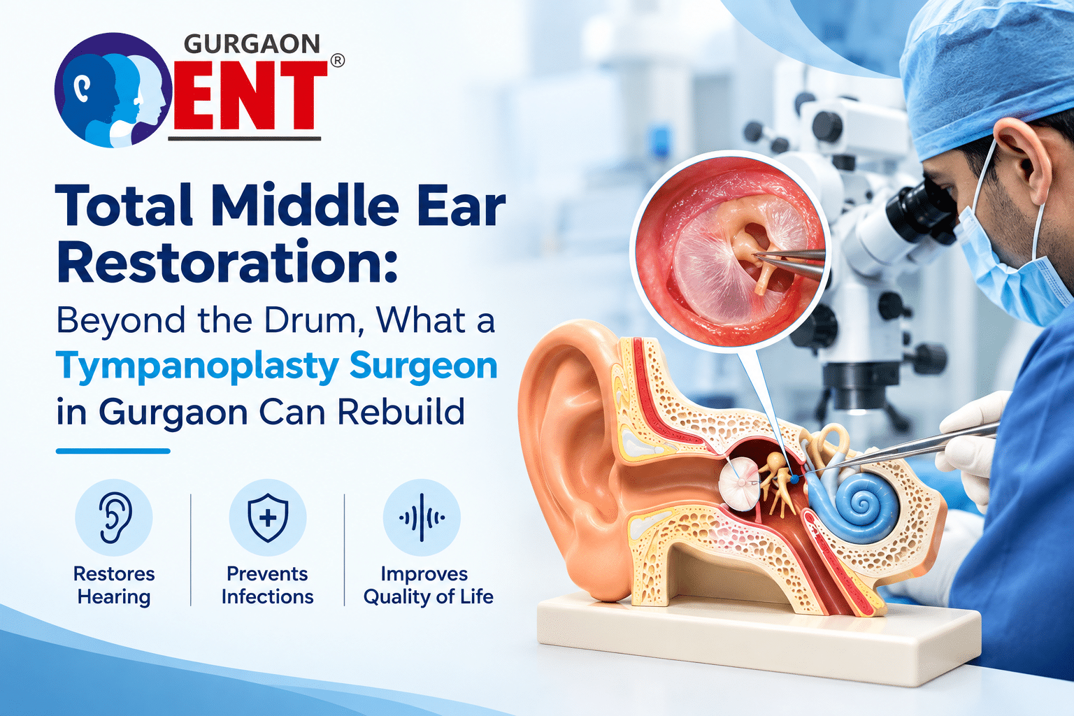
Say the word tympanoplasty and most people picture a patch stretched over a hole — which is fair, since that’s what a basic myringoplasty does. But the surgery, done properly, checks three separate structures: the eardrum itself, the chain of three bones behind it (malleus, incus, stapes), and the air-filled cavity they sit inside. Depending on the case, the mastoid bone behind the ear comes into the picture too.
Here’s the part that surprises most patients. A discharging ear left alone for years doesn’t just eat through the drum. It slowly corrodes the ossicular chain, and the long process of the incus goes first almost every time — it has the thinnest blood supply of the three bones, so it’s the one that gives out under sustained inflammation. Someone who’s been telling their doctor “I just need the hole patched” sometimes finds out on the operating table that half their hearing loss was coming from a snapped or dissolved incus, not the perforation they came in complaining about.
That’s the reasoning behind classifying these surgeries into types. Not paperwork for its own sake — the amount of rebuilding required swings wildly from one ear to the next.
Type I Through Type V: What Changes at Each Stage
Wullstein’s classification, still the reference point surgeons use today, ranges from Type I to Type V based on how much of the ossicular chain survives.
- Type I (myringoplasty): The three bones are intact and moving freely. Only the drum needs repair.
- Type II: Part of the chain — usually the malleus handle or an eroded incus — is gone, so the graft sits against whatever remnant is left.
- Type III: Enough of the chain has disappeared that the graft rests directly on the stapes head, effectively turning the drum itself into the sound conductor.
- Type IV and V: Reserved for extensive disease near the oval and round windows — often in ears that have already been operated on once and failed, or where infection has run for a very long time.
A related procedure, ossiculoplasty, rebuilds the chain using either the patient’s own repositioned bone or a prosthesis. Two terms surgeons throw around a lot here: PORP and TORP. A PORP (partial prosthesis) works when the stapes arch is still standing. A TORP (total prosthesis) is needed once that arch is gone entirely and the implant has to rest straight on the footplate. Both do the same job mechanically — turning what’s left of the chain into something closer to a single rigid piston carrying sound from the drum to the inner ear, a mechanism described in ossiculoplasty literature published through the National Institutes of Health.
Patients usually want one answer up front: will this fix everything in one sitting? That depends on which of these five types their ear actually falls into, and that’s not something an ear exam alone always settles — it often only becomes clear once the surgeon is under the microscope looking directly at the chain.
What the Graft Is Actually Made Of
Here’s something that catches people off guard: surgeons don’t reach for synthetic material to patch the drum. The standard choice is tissue harvested from the patient’s own head, usually one of two things — temporalis fascia (a thin sheet pulled from just above the ear) or tragal cartilage with its perichondrium (from the small flap of cartilage sitting in front of the ear canal).
Cartilage has been gaining ground for larger perforations and for ears where the Eustachian tube isn’t working well, since it holds its shape better than fascia and resists sinking back over time. One technique worth knowing about is the inlay “butterfly” cartilage graft, first described by Eavey in 1998 — a small button of tragal cartilage cut with a groove around its edge so it clips onto the perforation rim like a snap-fit part, detailed in published technical work on inlay cartilage tympanoplasty.
Placement matters too. Underlay puts the graft beneath the remaining drum tissue; overlay sits it on top; interlay wedges it between the drum’s two layers. Each comes with its own healing timeline and its own failure pattern — overlay grafts, for instance, carry a slightly higher risk of blunting at the front edge of the ear canal if not handled carefully. Which one gets used depends on where the perforation sits and how much healthy drum tissue is left to work with.
Endoscope or Microscope: The Debate Is Overstated
Patients ask about this more than almost anything else, usually because “endoscopic” sounds newer and they assume newer means better. It’s not that simple. Microscopic ear surgery has decades of track record and still does the heavy lifting when the mastoid needs to be opened up.
The endoscope earns its place elsewhere — it goes in through the ear canal on a thin camera, skips the incision behind the ear, and gives a wider view into corners like the sinus tympani or the front edge of a perforation that a microscope sometimes can’t see around. A study across three university hospitals covering 292 endoscopic ossiculoplasty cases recorded the average air-bone gap dropping from roughly 27 decibels before surgery to about 20 decibels afterward, with grafts holding in just over 94 out of every 100 cases.
A separate India-based comparison at a tertiary hospital, looking only at Type I procedures, found the endoscopic and microscopic groups performing about the same on hearing and graft outcomes. Neither wins outright. The choice hinges on where the perforation sits, whether the mastoid needs opening, and which technique the surgeon has done a thousand times rather than fifty.
The Damage That Happens Quietly, Year After Year
A discharging ear isn’t a cosmetic nuisance — it’s active inflammation, and inflammation is corrosive. The incus goes first, for the reason mentioned earlier, and that’s why patients who describe a decade of “just infections managed with drops” sometimes discover during surgery that the mechanical part of their hearing loss — a snapped bone, not a hole — was the bigger issue all along.
Delay compounds the problem. The longer chronic otitis media sits untreated, the higher the odds that a simple graft won’t be enough and full ossiculoplasty becomes necessary — or, in ears where a cholesteatoma has taken hold, revision surgery down the line.
What This Surgery Can Promise, and What It Can’t
Better to say this upfront than let someone walk out of the operating theatre expecting a miracle. The surgery aims to close the perforation, stop the discharge for good, and recover as much hearing as the remaining chain and inner ear allow. A clean Type I case with an intact chain tends to land close to normal hearing. A TORP or PORP reconstruction brings real improvement too, but a titanium or hydroxyapatite prosthesis, however well it’s seated, is a mechanical stand-in — it rarely matches the acoustic performance of the original three-bone chain working together.
Recovery isn’t a light-switch event either. It needs to keep ears out of the ear for some weeks, no travel in flights, hard nose blowing is not allowed, and keep checking the graft with a surgeon under a microscope. The condition of hearing can be evaluated only after two to three months, it can be done after the swelling of post surgery is completely cured and the real numbers settle in.
Why the Person Holding the Instrument Matters More Than the Instrument
Clinic marketing tends to lean hard on hardware — the newest endoscope model, the latest prosthesis alloy — as if the tool were doing the surgery. It isn’t. Deciding mid-operation whether a PORP or TORP fits better, judging how much cartilage a large defect needs, spotting an early cholesteatoma tucked behind a drum that otherwise looks fine — that judgment comes from repetition, not equipment. It’s built from having seen several hundred ears that all looked similar on paper and behaved completely differently once opened up.
That’s what an experienced Tympanoplasty Surgeon in Gurgaon brings that a brochure can’t: reading the specific anatomy in front of them and adjusting the plan mid-surgery when the ear doesn’t match what the pre-op scan suggested.
Signs This Surgery Is Worth Discussing
- Repeated ear discharge that flares up after swimming, showering, or a common cold
- Conductive hearing loss where a hearing aid feels like patchwork rather than a fix
- A “dry” perforation that still comes with noticeably reduced hearing — often a sign the chain, not just the drum, is involved
- Years of managing chronic otitis media with ear drops alone, without ever addressing what’s happening structurally
- A diagnosis of early cholesteatoma, which needs surgical attention regardless of how much hearing is currently affected, since it keeps eroding bone in the background
Aftercare Decides How Long the Result Lasts
Modern graft closure rates sit comfortably above 90 percent in the studies cited above, but “closure” only means the hole sealed shut. Whether hearing stays stable for years afterward comes down to Eustachian tube function, how well future infections are kept away, and whether a prosthesis stays seated where it was placed. Follow-up visits exist to catch a retracting graft or a shifted prosthesis early, while it’s still a five-minute fix rather than a second surgery.
Frequently Asked Questions
Is tympanoplasty only about closing a hole in the eardrum?
No, that’s myringoplasty. Full tympanoplasty also checks and, where needed, rebuilds the three bones behind the drum, since chronic infection tends to damage both together.
How soon will I know if my hearing has actually improved?
Initial healing runs a few weeks, but the audiogram that tells you the real result is usually done two to three months later, after post-surgical swelling has fully settled.
Will I still need a hearing aid afterward?
Often not, especially with a straightforward perforation and an intact chain. In more involved reconstructions, some residual loss can remain — your surgeon should walk you through that possibility before surgery, based on what’s actually found in your ear.
Is endoscopic surgery always the better option?
Not automatically. Outcomes run close between the two approaches in most published comparisons. What matters more is your specific anatomy and how extensive the disease is — not which method sounds more current.
What happens if a long-standing perforation is just left alone?
Ongoing discharge keeps eroding the ossicular chain in the background and raises the odds of complications like cholesteatoma. Getting it looked at earlier tends to mean a simpler surgery, not a more complicated one.
The Short Version
The drum gets the attention because it’s the part people can picture. What actually determines the hearing outcome is everything sitting behind it — three tiny bones and the space they occupy. If a “small hole” has been on your list for years, or the hearing loss you’re dealing with doesn’t quite match what a simple perforation should cause, a proper microscopic exam is the only way to know what’s actually going on.
Dr. Ravinder Gera at Gurgaon ENT Clinic has worked through this range himself — from routine myringoplasties to more involved ossicular rebuilds — and prefers to lay out exactly what a given ear needs before recommending anything. As a practising Tympanoplasty Surgeon in Gurgaon, his approach starts with what the ear shows on examination, not with a fixed technique applied to every case that walks in.
If a long-standing perforation, chronic discharge, or unexplained hearing loss has been on your to-do list, Gurgaon ENT Clinic is at 112, Apna Bazar, Gurudwara Road, Gurgaon. Call +91-9810340495 to book an evaluation.
Recent Post
-
Total Middle Ear Restoration: Beyond the Drum, What a Tympanoplasty Surgeon in Gurgaon Can Rebuild
September 9, 2026
-

Globus Sensation: Understanding the “Lump in Throat” Feeling With an ENT Specialist in Gurgaon
September 7, 2026
-

Cognitive Hearing Science: How a Hearing Specialist Doctor in Gurgaon Prevents Brain Fog
August 24, 2026